
Kaleigh has several fantastic studies that are in the publication process. So we are going to call 2024 the “Year of Connors et al”. She has worked really hard over the past couple of years to develop several novel experimental systems for use in our lab. The first one is the rat brain slice culture system that she validated and adapted for our use to study neurotropic bunyaviruses.
Journal of General Virology: Acute Rift Valley fever virus infection induces inflammatory cytokines and cell death in ex vivo rat brain slice culture
I posted about it on Twitter (X) which you can read about here.
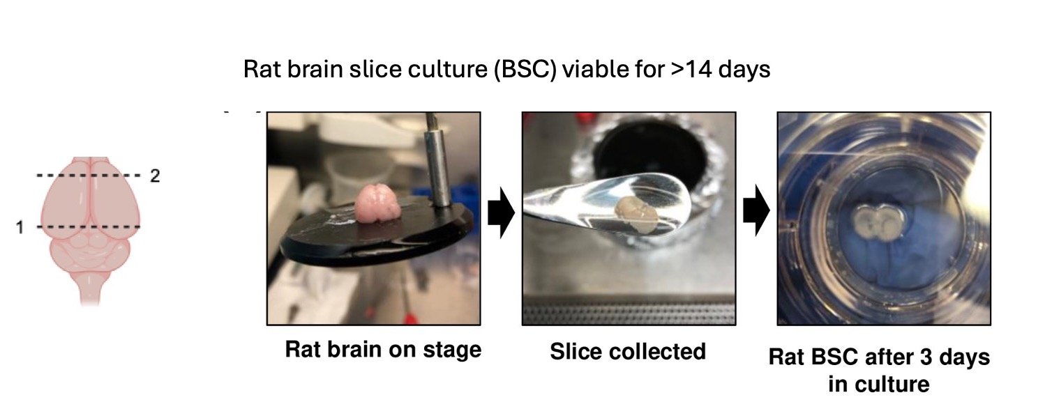
What continues to amaze me about this is the fact that these slices are viable for at least 14 days, and Kaleigh did a lot of validation to understand what is happening in the cultures over time. In fact, we use the slices for infection studies after they have been in culture for 10 days because this give the tissue time to calm down from the trauma of slicing. The brains are from 6 day old rat pups, which is why they are able to stay viable for a long time. Similar slices from adult animals would likely not stay viable.
We plan to use these to test therapeutics in brain slices prior to in vivo testing. We hope this gives us a model that is intermediate between immortalized cell lines and cumbersome in vivo BSL-3 studies.
Kaleigh also produces the most beautiful fluorescent images of anyone in the lab (**Zach can certainly rival her for primary neuron images!). Look at these images from cleared slices. Nuclear filaments with active caspase 3 are always striking.

Using the vibratome to slice the brains is pretty cool. Here are some photos of a brain stabilized in agar and then mounted on a cork (See, the PI’s wine consumption comes in handy!). Also, a photo of Zach learning how to slice the brains.


