
Extra! Extra! Hear all about it! Our premier post-doc Dr. Rachael Rush has a new publication in the Journal of General Virology! Entitled, “Rift Valley fever virus infects trophoblast cell lines in an LRP1-dependent manner,” the paper suggests LRP1 to be a determinant of RVFV infection at the maternal-fetal interface as evidenced by RVFV’s ability to replicate within human trophoblast cell lines efficiently. This publication ALSO happened to be one of the most read papers in the Journal of General Virology in the past month!
Immortalized human trophoblast cell lines can provide insight into the mechanisms of cellular infection at the maternal-fetal interface. JEG-3 and JAR cells provide a model cell morphologically similar to cytotrophoblasts while the HTR-8/SVneo (HTR-8) cell line exhibits extravillous morphology which provides a useful analog for syncytiotrophoblast cells along the uterine wall. We have previously demonstrated evidence of the ability of RVFV to infect and replicate within both cytotrophoblasts and syncytiotrophoblasts of primary human placenta explants. The work of this paper uses the aforementioned cell lines to elucidate how RVFV infection is mediated within these cells and provide perspective on the mechanisms of RVFV vertical transmission and infection of the placenta.
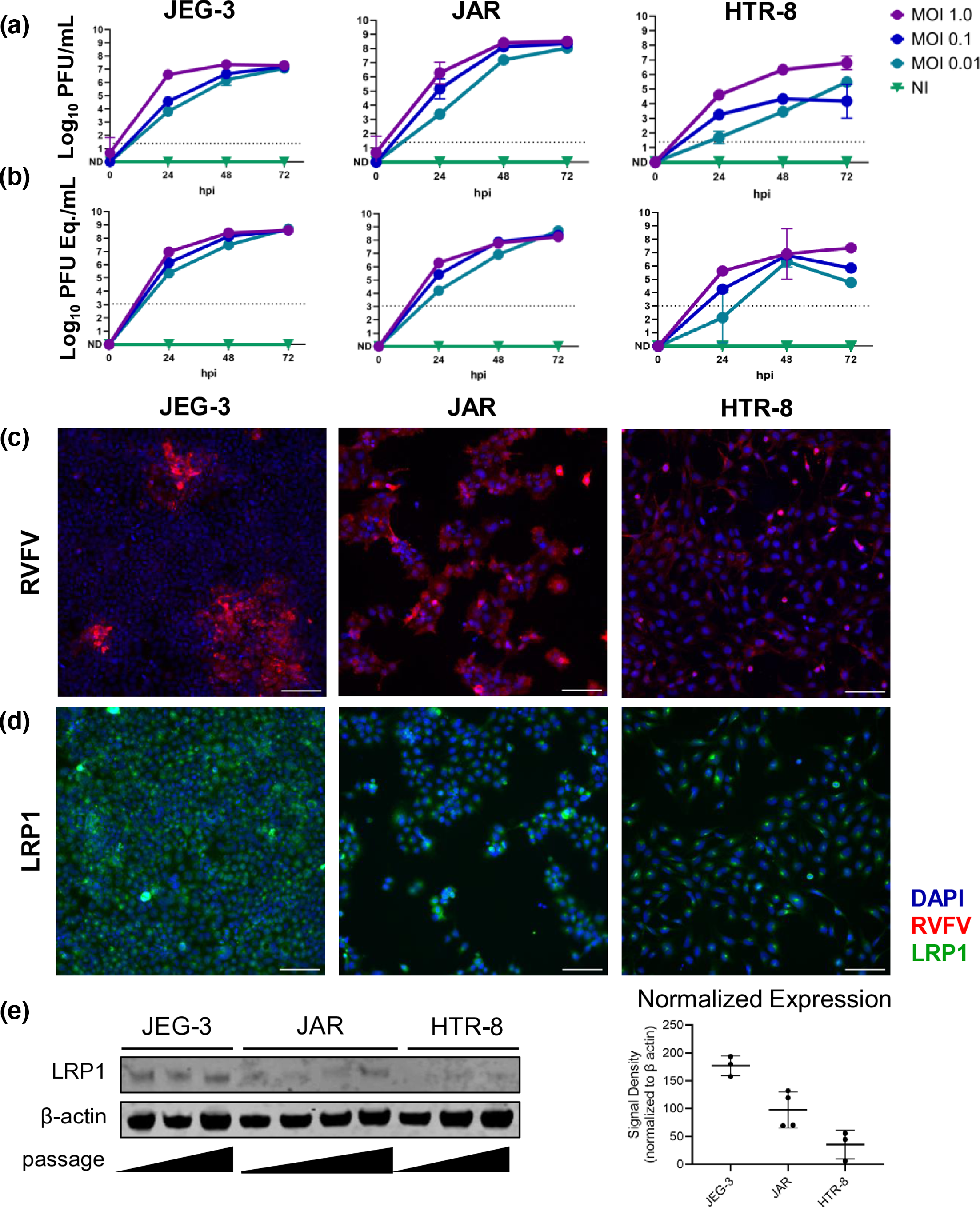
In vitro infections of JEG-3, JAR, and HTR-8 cells at MOIs of 0.01, 0.1, and 1.0 all showed evidence of RVFV infection and the expression of LRP1. HTR-8 appeared to be less permissive of RVFV infection when examining vRNA as a demonstration of infectious virus within the system. This was supported by immunofluorescent staining that showed less RVFV antigen staining in HTR-8 cells than the JEG-3 and JAR counterparts at 24 hours post infection (Figure 1C). This coincides with the expression of LRP1 within these cell lines, in which HTR-8 cells are shown to exhibit comparatively less LRP1 through immunofluorescent staining as well as western blot.
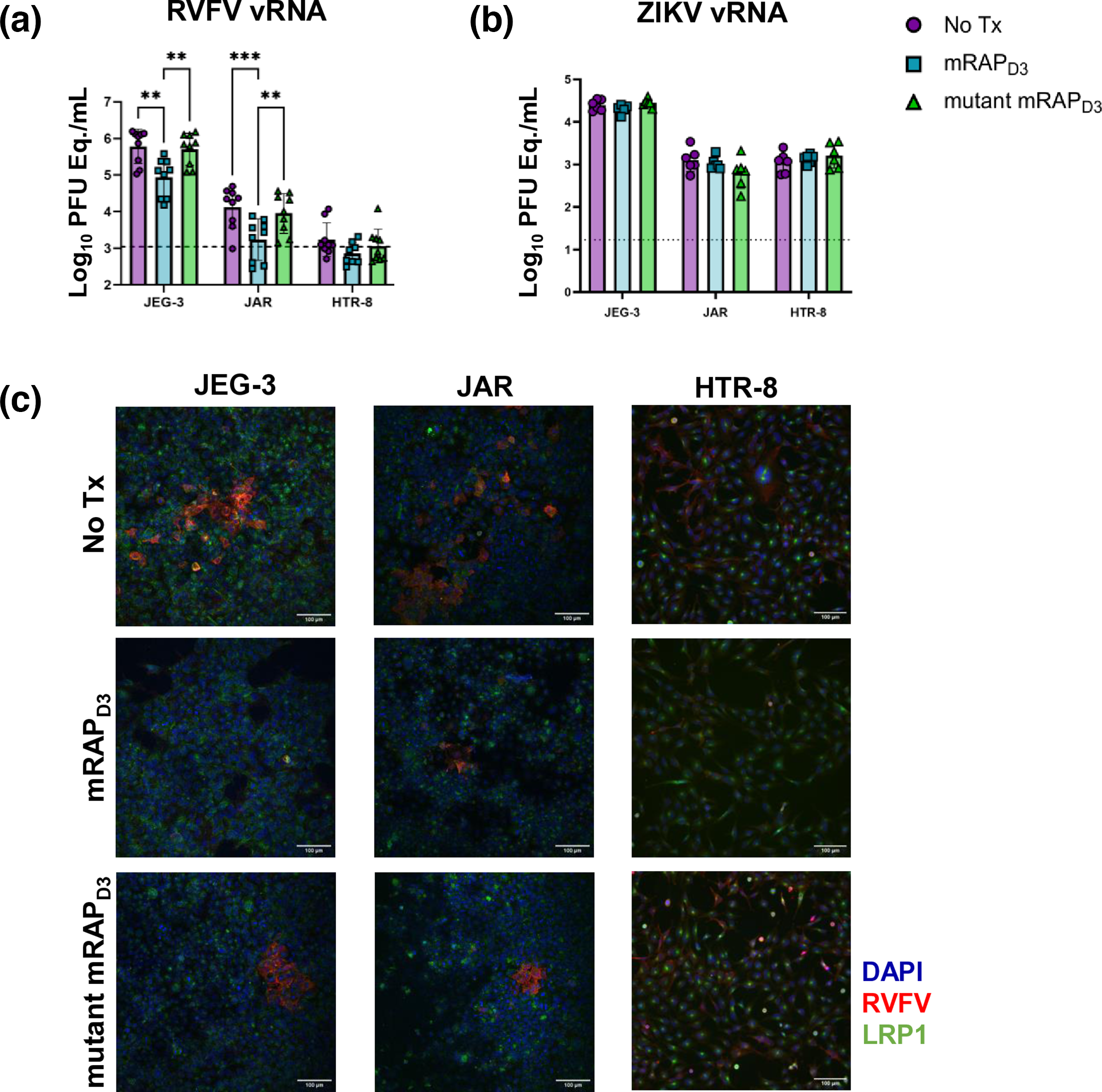
In order to further understand the dependency of LRP1 in RVFV of these trophoblast cell lines, we used receptor-associated protein (RAP) as a competing ligand to bind to LRP1, blocking the interaction of RVFV with LRP1 on the surface of these cells. Treatment of cells with RAP reduced RVFV viral titres in both JEG-3 and JAR cells, with the vRNA levels of HTR-8 cells already being so close to the limit of detection that significant difference could not be discerned. This was not true of cells treated with RAP before infection with Zika virus (ZIKV), a flavivirus not previously known to interact with LRP1 or similar LDLRs. Immunofluorescent staining also demonstrates this relationship between RAP treatment and decreased presence of RVFV within the cells (Figure 2C). This suggests that the results showing the reduction in vRNA and antigen staining is specific to the function of RAP treatment as a competitive inhibitor of LRP1.
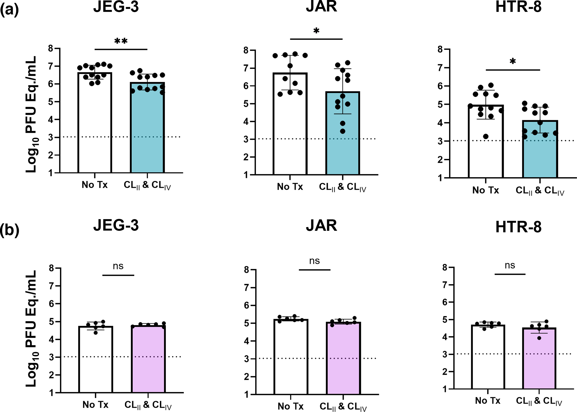
The specific RAP used for these treatments is Murine RAP domain 3 (mRAP D3) which binds to all LDLRs, not solely LRP1. In order to examine the role of LRP1 inhibition more exclusively, we utilized recombinant LRP1 decoy proteins for the extracellular domain clusters II and IV (CLII and CLIV) fused to a human Fc. When pre-treating RVFV inoculum with these CLII and CLIV decoy proteins it resulted in a significant reduction of vRNA and relative infectivity in all cell lines when compared to control-treated cells. This relationship was not seen when ZIKV inoculum was treated the same way. These results support the notion that RVFV requires binding to CLII and CLIV, and the blocking of that interaction reducing infectivity indicates that LRP1 is used by RVFV to enter and infect these trophoblast cell lines.
Outstanding work by Rachael and all who contributed! LRP1 and RVFV infection become better understood every day because of hard work like this!