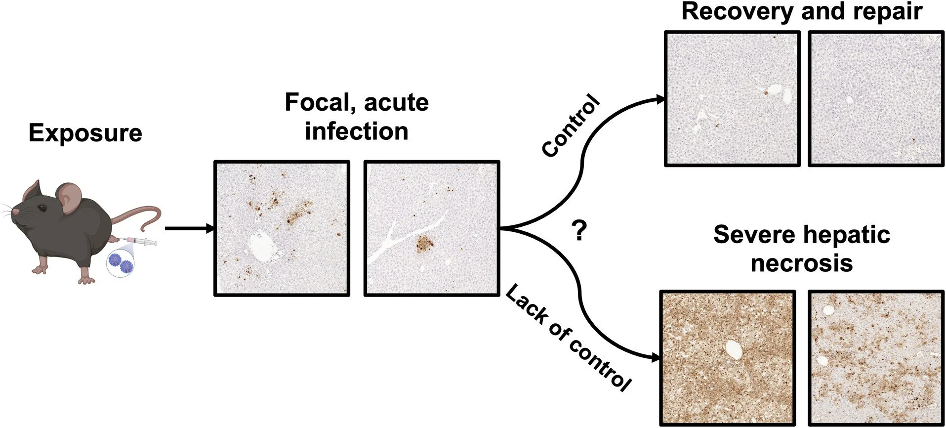
It’s been a busy summer here at the Hartman lab. Among the many projects going on currently, we have a new Sterling et al. publication in the Journal of Virology to show for all of this hard work!
Published earlier this summer, “Oropouche virus causes acute hepatitis in mice controlled by type I interferons,” presents two models of Oropouche virus hepatic disease in mice. Fatal cases of OROV in humans have been attributed to severe coagulopathy within the liver, as well as mild liver injury for those with mild cases of Oropouche fever. However, the role of the liver in the pathogenesis of OROV is still rather unexplored.
Cade has developed a lethal model of OROV infection that causes severe liver necrosis similar to what is seen in fatal cases of OROV infections in human, as well as a non-lethal model of self-resolving acute hepatitis that serves as an analog for a milder disease case. Both models cause focal hepatic cell death in mice which can progress to more severe liver necrosis and death when Type I interferon receptors are blocked. These models can serve as valuable tools for the preclinical evaluation of OROV vaccine platforms and therapeutics as well as a means of understanding how OROV causes liver damage.
In Cade’s non-lethal model, immunocompetent C57BL/6 mice were infected with the prototypical OROV strain BeAn19991 via the footpad. These mice were euthanized at 1, 3, and 5 days post-infection for their tissues to be harvested for quantification of viral load and histology (Figure 1A). There was no infectious virus found in the spleen, kidney, lung, or brain at any of the euthanasia time points. Meanwhile, the livers of infected mice had detectable levels of OROV as early as 1 day post-infection (dpi), with the viral titers peaking at 3dpi (Figure 1D).
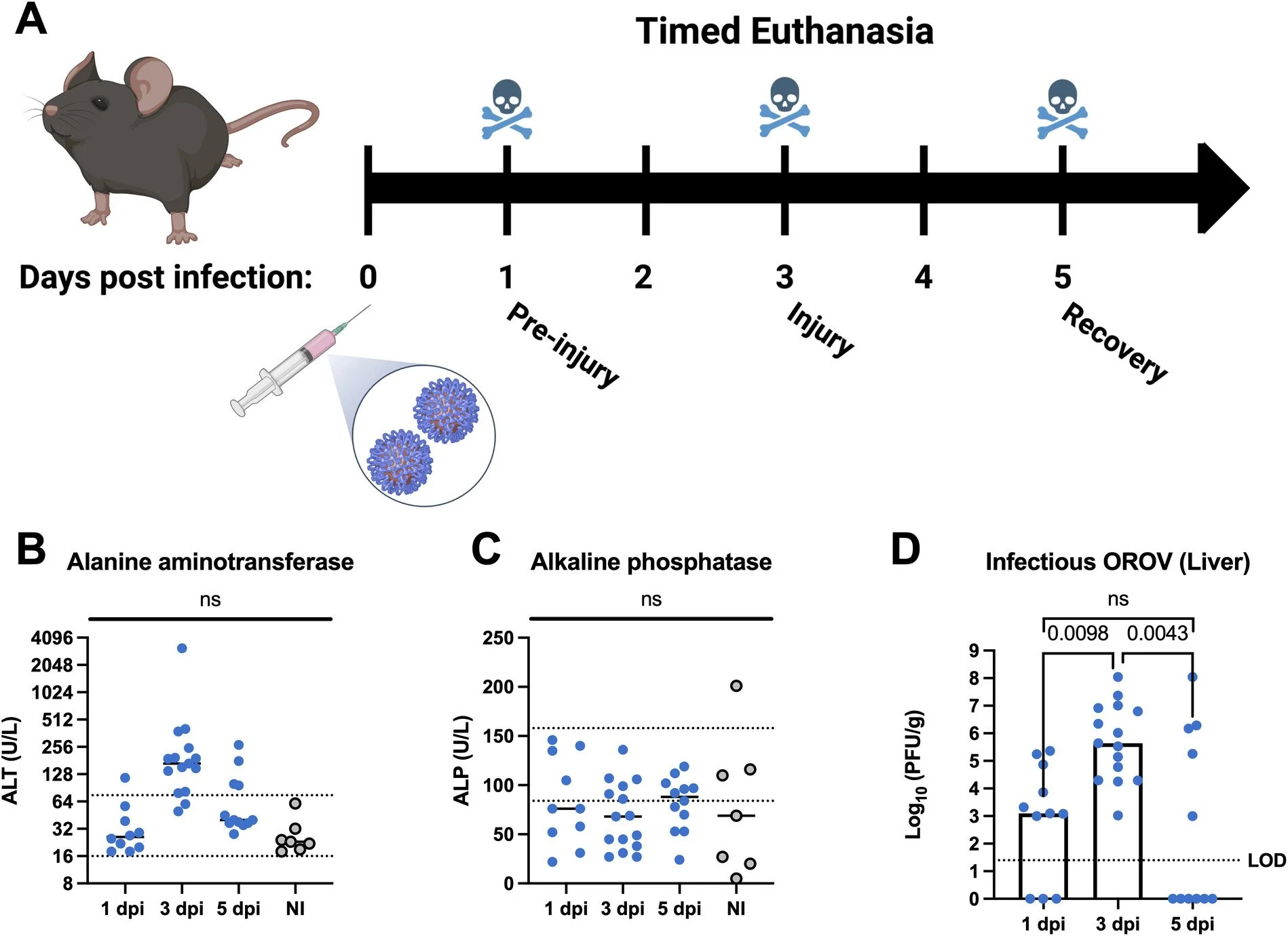
The presence of virus in the liver along with elevated liver enzymes at 3dpi (Figure 1B) suggested there was damage in the liver, so histopathology was performed. The beautiful images shown below were obtained through hematoxylin and eosin (H&E) and TUNEL staining, and they reveal OROV-induced necrotic and inflammatory areas across the livers of infected mice.
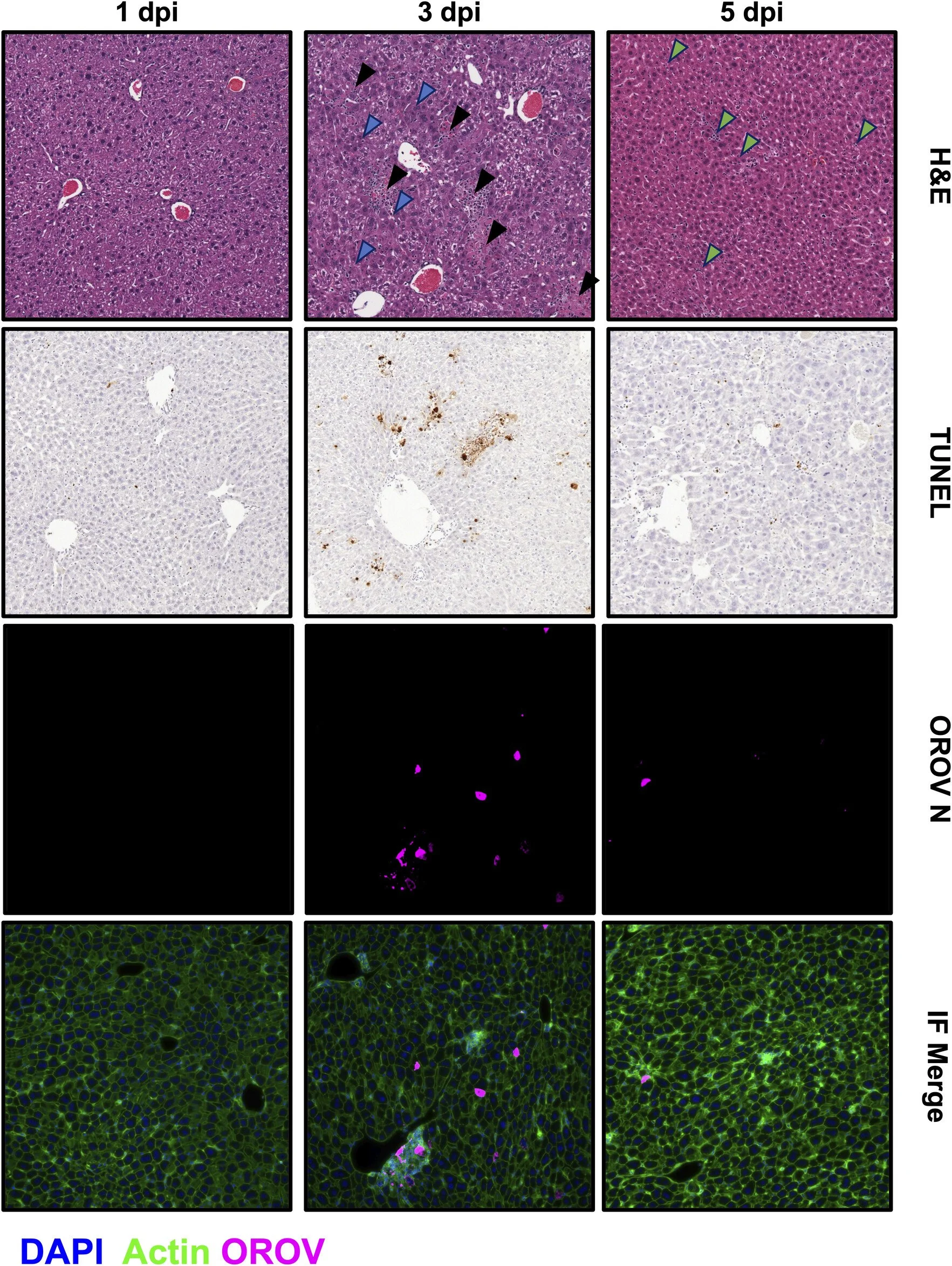
Cade’s exceptional liver-imaging capabilities help paint the picture of how OROV infection progresses through the liver over time. Histopathological damage induced by the infection increases from 1dpi to 3dpi, before showing signs of recovery and repair by 5dpi through the visible lack of focal damage or accumulation of immune cells.
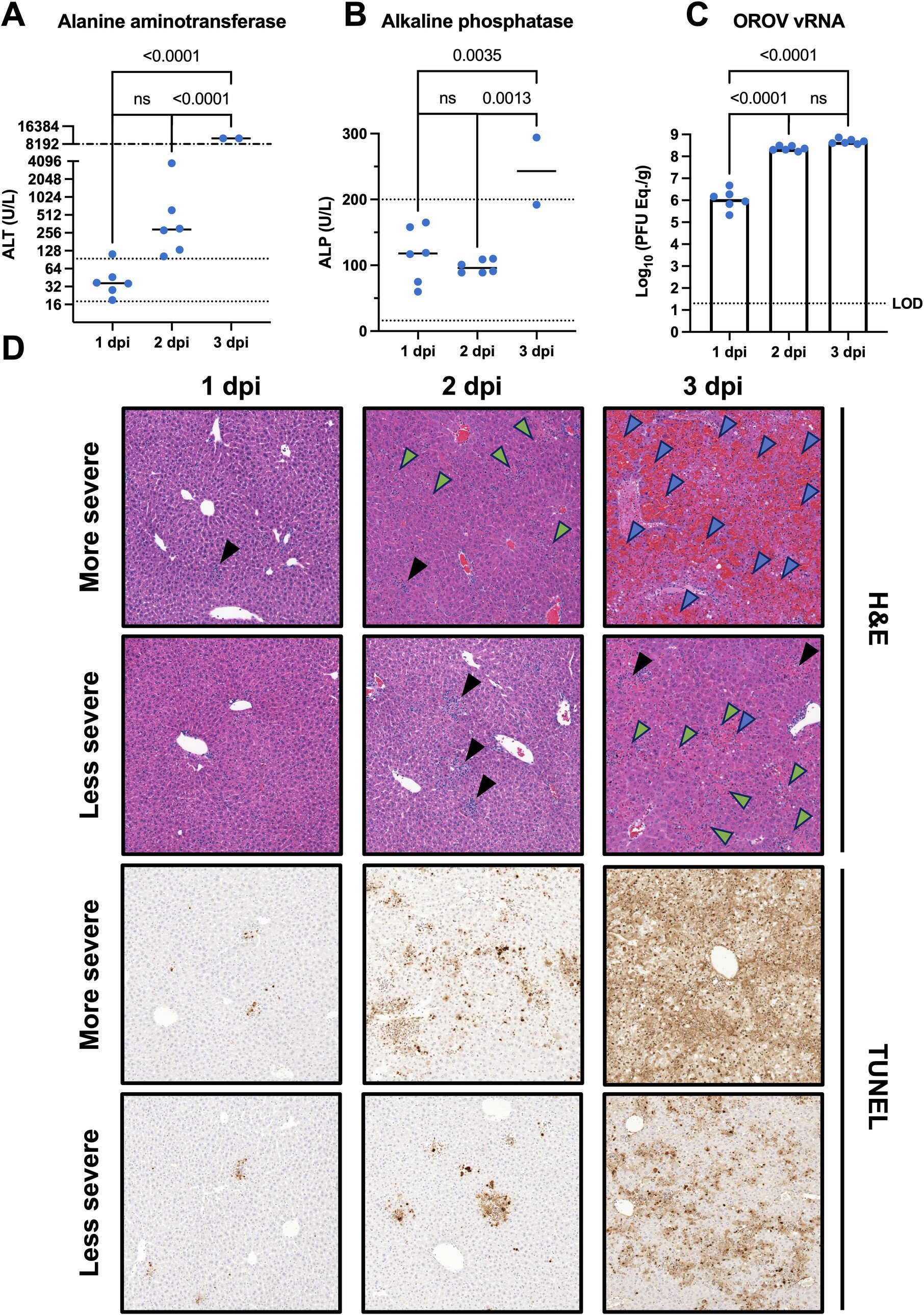
Cade’s lethal model of OROV infection utilizes a MAR1-5A3 clone monoclonal antibody to bind the Type I IFN receptor complexes and immunocompromise the mice by preventing recognition of Type I interferons. Treating mice with MAR1 prior to OROV infection resulted in near-uniform lethality at the highest dose by 5dpi. Severe disease and liver damage were seen in these mice which prompted a timed euthanasia study to assess viral titers, blood chemistry, and histopathology. This timepoint study revealed the rapid onset of hepatic damage. Necroinflammatory foci, necrosis, and hemorrhage are severe by 3dpi (Figure 6D). The deficient immune response caused by the presence of MAR1 prior to OROV infection shows a course of disease in which OROV can overwhelm the tissues of these mice and cause severe hepatic disease, necrosis, and death. In both the sublethal and lethal model the elevated liver enzymes and similar histopathology by 2dpi suggest a similar disease progression. However, the ability of the murine immune system to mount a sufficient response seems to determine whether the infection resolves and the liver repairs, or the more severe outcomes prevail.
A more contemporary OROV outbreak isolate (CDC240024) was also used with the MAR1 lethal model, causing markedly less lethality than the historical prototype strain (BeAn19992). Histopathological outcomes were similar, although the clinical window was compressed and the time to death was delayed by approximately one day. This may suggest current human isolates may be less adapted to infecting laboratory mice as opposed to a strain with a rich passage history such as BeAn19991. Together, these lethal and sublethal models of OROV infection paint the picture of how OROV hepatic disease progresses within immunocompetent and immunocompromised mice. This work prompts further investigation into the mechanisms which determine whether or not OROV will be controlled by the immune response within the liver or progress into severe hepatic necrosis. Stellar work, Cade!